Alzheimer’s May Shrink Two Brain Regions Years Before Memory Loss, Study Finds
Brain scans from Johns Hopkins scientists show two regions tied to memory and emotion shrink years before the hippocampus, offering an early warning sign for Alzheimer’s risk linked to tau protein buildup.
In this piece
Johns Hopkins researchers have discovered that two brain regions involved in memory and emotion begin shrinking up to a decade before Alzheimer’s memory loss…
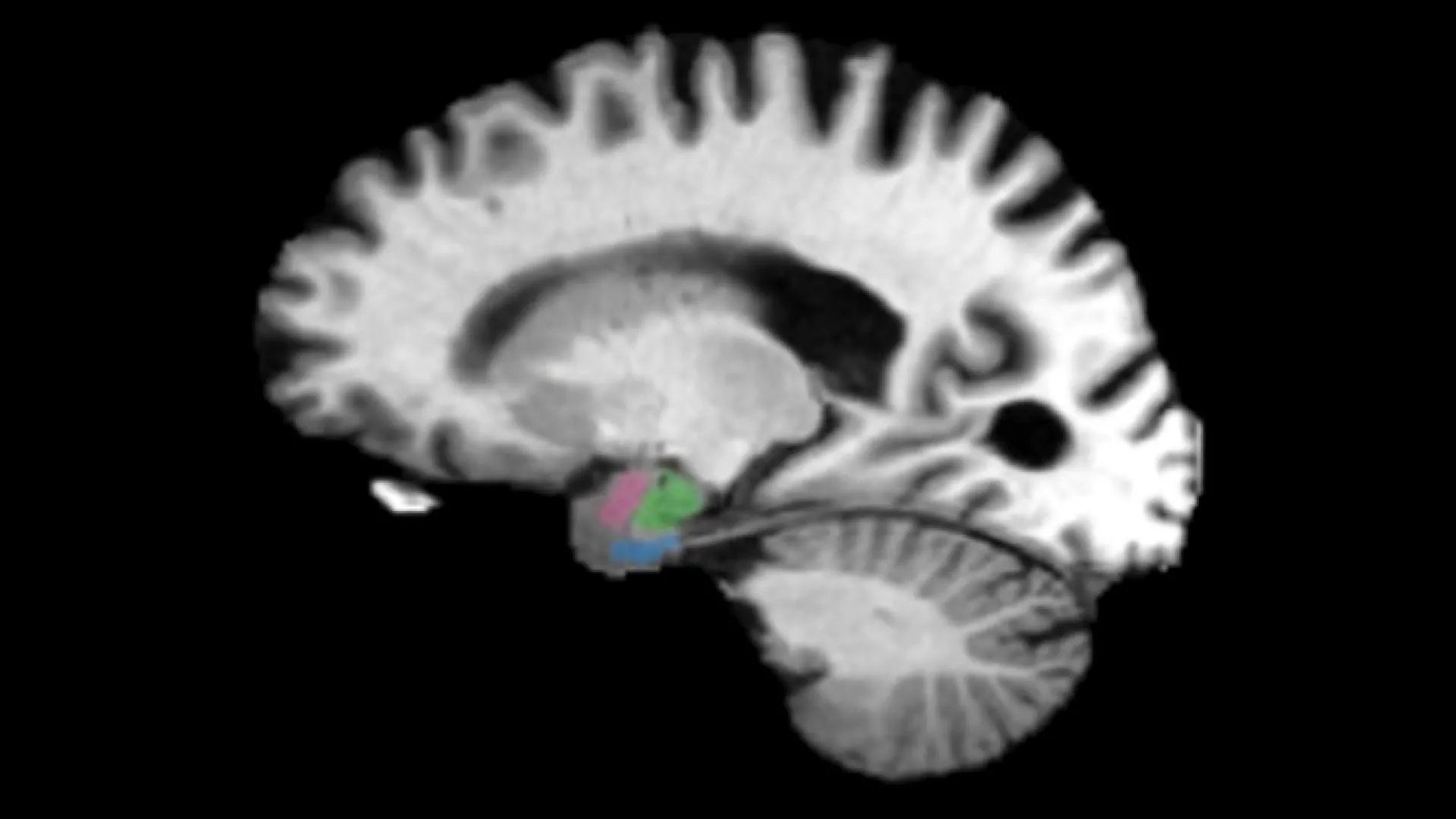
Researchers at Johns Hopkins have identified potential early structural markers of Alzheimer’s disease that can emerge up to a decade before clinical memory loss becomes evident. Advanced neuroimaging indicates that two distinct brain regions responsible for memory and emotional processing begin to atrophy well before measurable shrinkage appears in the hippocampus, the structure traditionally monitored by clinicians.
In current diagnostic practice, physicians often track the volume of the hippocampus to gauge the onset and advancement of neurodegenerative decline. Because the hippocampus plays a central role in forming and consolidating new memories, noticeable loss of volume in this area has long served as a primary hallmark of progressing Alzheimer’s disease.
The new imaging investigations reveal that subtle atrophy develops earlier in two interconnected regions tied to emotional regulation and memory processing. Researchers determined that these early volumetric reductions closely correlate with the abnormal accumulation of tau protein, one of the primary pathological drivers associated with the disease.
Alzheimer’s disease is widely characterized by the progressive accumulation of toxic protein deposits in the brain, notably beta-amyloid plaques and neurofibrillary tangles composed of tau. While amyloid accumulation often begins silently years before diagnosis, the spread and clustering of tau protein are closely coupled with direct cellular damage, synaptic disruption, and tissue loss.
A persistent obstacle in addressing Alzheimer’s disease has been the delayed detection of cellular damage, as conventional clinical assessments frequently occur only after irreversible cognitive impairment has set in. When individuals first present with overt confusion or memory lapses, substantial neuronal networks have typically already experienced significant deterioration.
Detecting physical brain shrinkage years before clinical symptoms manifest opens a critical window for medical intervention. Identifying measurable structural changes in vulnerable regions during this asymptomatic phase could enable healthcare providers to categorize individuals at elevated risk far sooner than standard diagnostic thresholds currently permit.
Early identification is increasingly vital as medical researchers and pharmaceutical developers introduce therapies intended to slow or halt neurodegenerative disease progression. Interventions designed to clear abnormal protein accumulations or protect neuronal integrity are broadly considered most effective when initiated prior to widespread structural loss in the hippocampus and surrounding cerebral cortex.
Integrating sensitive scans focused on these early-shrinking areas could ultimately reshape how clinical trials select participants and measure therapeutic success. By tracking volume changes alongside tau accumulation, clinical investigators can better evaluate whether experimental treatments successfully shield vulnerable neural architecture from progressive decay.
While these imaging observations mark an important step forward, broader validation across diverse patient cohorts will be essential to establish standardized clinical protocols. Researchers will need to determine how reliably these localized volumetric measurements predict individual cognitive trajectories and how accessible such neuroimaging methods can become in routine medical care.
Primary source: ScienceDaily. Newzly rewrites and structures source reporting for clarity; verify critical details at the original.